Muscle Function, Diet, and Metabolism in Active Horses

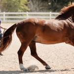
All movement in the horse—from the explosive power of the jumper and cutting horse to the slow, steady trot of the endurance horse—depends on the contraction of skeletal muscles. The horse’s muscles function in several different ways depending on their particular characteristics and the fuel they derive from forages and concentrates in the horse’s diet.
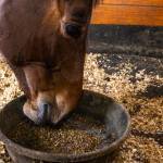
When a horse exercises, its muscles use small molecules called ATP (adenosine triphosphate) to stimulate muscle contraction. Horse muscle has very little stored ATP, but it has numerous metabolic pathways that can produce ATP as quickly as it is used. These pathways use several fuels to produce ATP, and one objective of feeding an exercising horse is to optimize the stores of these fuels so the muscle can continue to contract without fatiguing. The most important fuel for active horses is glycogen (a string of glucose molecules), which is stored predominantly in the muscle and to a lesser degree in the liver, and fat, which is stored mostly in adipose tissue and to a lesser degree in the muscle. During hard exercise such as a race, muscle glycogen is the major fuel used by the horse, but fat is important to supply calories for lower intensity training and to meet the horse’s maintenance energy requirement.
Skeletal muscle consists of bundles of long spindle-shaped cells called muscle fibers that attach to bone by tendinous insertions. The blood vessels and nerves that nourish and control muscle function run in thin sheets of connective tissue that surround bundles of muscle fibers. Each nerve branch communicates with one muscle fiber at the motor end. The nerve and all the muscle fibers that it supplies are together termed a motor unit. Each time a nerve is stimulated, all of the muscle fibers under its control will contract.
A muscle’s unique ability to contract is conferred by the highly organized parallel and overlapping arrangement of actin and myosin filaments. These repeating contractile units or sarcomeres extend from one end of the cell to another in the form of a myofibril. Each muscle fiber is packed with myofibrils that are arranged in register, giving skeletal muscle a striated (marked by parallel stripes or grooves) appearance under the microscope. Muscle contraction occurs when the overlapping actin and myosin filaments slide over each other, serving to shorten the length of the muscle cell from end to end and mechanically pulling the limb in the desired direction. The sliding of the filaments requires chemical energy derived from elements of the horse’s diet.
Different forms of myosin (isoforms) can be found in different muscle fibers and these different isoforms affect the speed with which a muscle cell can contract. In addition, fibers with specific contractile speeds will also possess characteristic metabolic energy producing capacities. Type 1 fibers contract slowly, are ideally suited for endurance, and are able to hold a tetanic twitch for long durations without fatigue. Their resistance to fatigue is in part related to their high density of mitochondria, which confer a high aerobic or oxidative capacity. In addition to having the highest oxidative capacity, type 1 fibers also have the highest lipid (fat) stores, the highest density of capillaries, the lowest glycogen stores, and the lowest glycolytic enzyme capacity of the three fiber types.
Fast-twitch muscle fibers or type 2 fibers are readily divided in the horse into type 2A and 2B fibers. Type 2B fibers have the fastest contractile speed, the largest cross-sectional area, the highest glycogen stores and glycolytic capacity, and the lowest oxidative capacity. As such, they are ideally suited to short fast bursts of power. Type 2A fibers are intermediate in contractile speed and metabolic properties between type 1 and type 2B fibers. Most large muscles in the horse contain a mixture of muscle fiber types.
The muscle fiber composition, the percentage of type 1, 2A and 2B fibers, and muscle fiber crosssectional areas vary greatly between muscle groups, among individual horses, and between breeds. These proportions are not constant, as training can alter the fiber composition and fiber size in the same muscle over time. Propulsive locomotor muscles, such as the gluteus, contain a predominance of fast-twitch type 2 muscle fibers, with the highest density of type 1 fibers located deeper within the muscle.
In general, Quarter Horses and Thoroughbreds have the highest percentage of fast-twitch muscle fibers, 80-90%; Standardbreds have an intermediate number, 75%; and donkeys have the lowest percentage of type 2 fibers in locomotor muscles. With growth and training, there is a change in the length and breadth of a fiber, and a change in the proportion of fiber types rather than an increase in the number of muscle fibers. In young horses intensively trained at speed, the proportion of type 2A fibers increases concomitant with a decrease in type 2B fibers.
When a muscle contracts during exercise, it does so in response to a predetermined recruitment of particular muscle fibers. This orderly recruitment of muscle fibers leads to smooth, coordinated movement. As exercise begins, a select number of motor units, each of which contains fibers of the same type, are recruited to provide the power to advance the limb. At slow exercise intensities, type 1 muscle fibers and a small number of type 2A muscle fibers are stimulated. The force produced by any muscle is proportional to the cross-sectional area that is active. As the speed or duration of exercise increases, more muscle fibers will be recruited and this occurs in the order of their contractile speed. Only at near-maximal exercise intensities or after several hours of submaximal exercise are type 2B fibers recruited.
More than dietary energy is necessary to keep horses moving smoothly as they train and perform. Electrolytes such as calcium are important in the function of nerves and muscles. When a nerve is stimulated, a wave of electrical depolarization occurs that quickly reaches the neuromuscular junction. In response, the nerve terminal releases acetylcholine, which binds to the motor-end plate of the muscle fiber and initiates electrical depolarization of the muscle cell membrane. Electrical depolarization of the muscle cell membrane triggers the release of calcium that is sequestered in intracellular membranous storage sites (sarcoplasmic reticulum) into the myoplasm via the calcium release channel. Increased concentrations of intracellular calcium allow the interaction of actin and myosin filaments, which then slide over each other in a ratchet-like fashion to produce a contraction (cross-bridge cycling). Tension is generated as the shortening filaments tug both ends of the myofiber toward the middle. Muscle must relax after each contraction by actively pumping calcium back into the storage sites, preventing actin-myosin interaction. In addition, ion pumps in the cell membrane actively repolarize the muscle cell membranes. All of the processes necessary for relaxation are active, meaning that they require energy in order for them to occur.
Information in this article was taken from Advances in Equine Nutrition.
Most Popular
 Putting Weight on a Skinny Horse (326,413)
 Benefits of Beet Pulp for Horses (217,008)
 Hot Blood, Warm Blood, Cold Blood in Horses (174,279)
 Possible Link Between Selenium and Cribbing in Horses (163,717)